Life Cycle: "Salivarian pattern: Trypomastigote forms circulate within the blood of a vertebrate host. They are extracellular parasites. A blood-feeding vector such as a tse-tse fly ingests trypomastigote along with its blood meal. Trypomastigotes continually divide by fission within the midgut over a period of 1-2 weeks. Over the next week they migrate anteriorly in the gut, eventually entering the salivary gland and accumulating there. Within the salivary gland the parasite transforms into an epimastigote form and continues to divide. They eventually transform into a metacyclic trypomastigote and are now infective to another vertebrate host. After inoculation into the blood stream of a new vertebrate host the parasite continues to divide in the trypomastigote form.
Stercorarian pattern: Trypomastigote forms circulate within the blood of a vertebrate host. They are extracellular parasites. A blood-feeding vector such as a bed bug ingests trypomastigote along with its blood meal. Trypomastigotes transform into epimastigotes within the midgut After about 1 week they transform into metacyclic trypomastigotes. When the host feeds, it often defecates at the same time, releasing metacyclic trypanosomes that contaminate the wound. Within the vertebrate the trypomastigotes are phagocytosed by host defense cells or they may enter other cells such as cardiac muscle. During this intracellular phase of existence they transform into amastigotes and multiple by fission. Large numbers of amasitgotes accumulate and form pseudocysts. Eventually the amastigotes transform into trypomastigotes, rupture the host cell, and enter the circulation. They may be ingested by a vector or infect a new host cell, again becoming amastigotes and repeating the cycle" (University of Alberta).
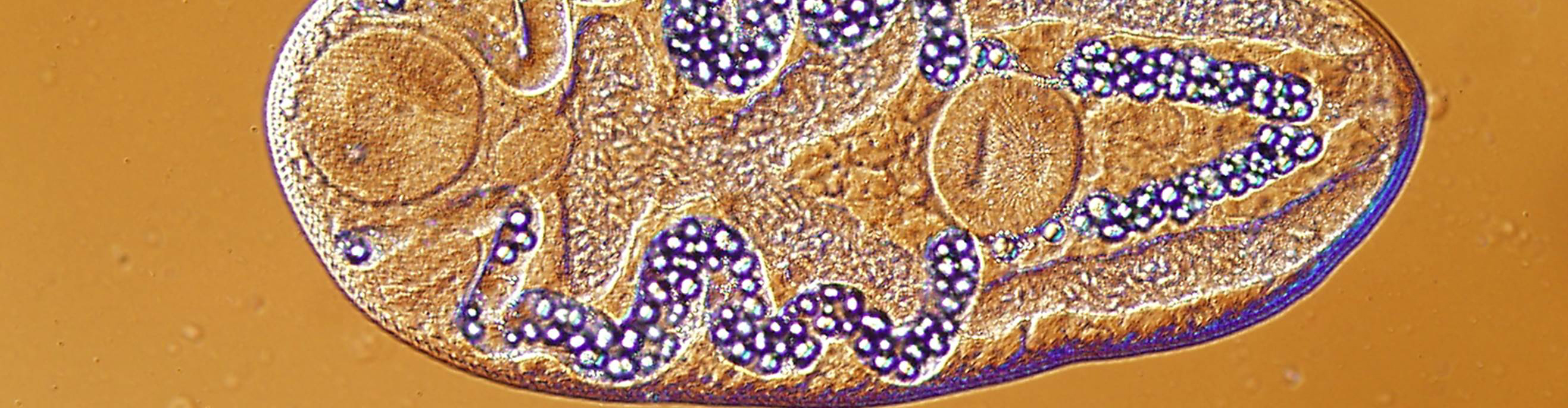
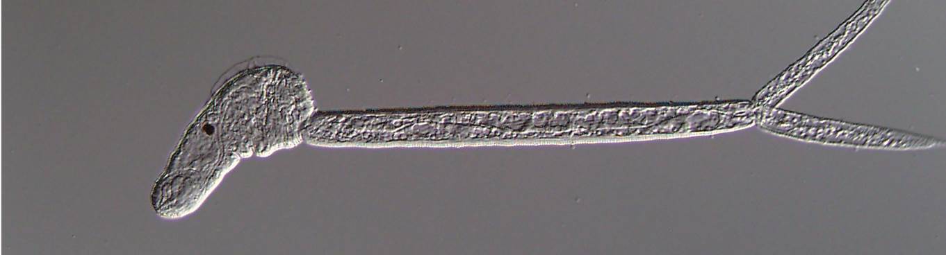
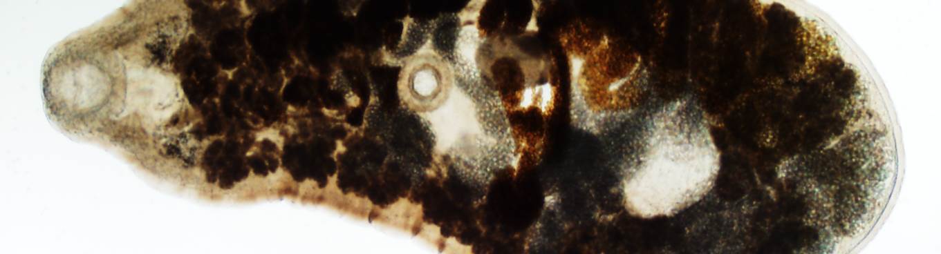
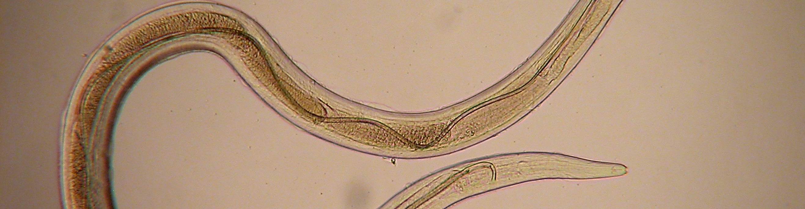
Description:
The body varies in general shape, some thin and some stout specimens being found. Nucleus, in most cases, is placed with its long axis parallel to the long axis of the body. The parabasal body is elliptical and is usually perpendicular to the long axis of the body. Undulating membrane is broad. Some specimens showed vacuoles scattered through the cytoplasm of the body.
Sources:Roudabush, R.L. and Coatney, G.R. 1937. On some blood protazoa of reptiles and amphibians. Transactions of the American Microscopical Society, Vol. 56, No. 3, p. 291-297.
University of Alberta.