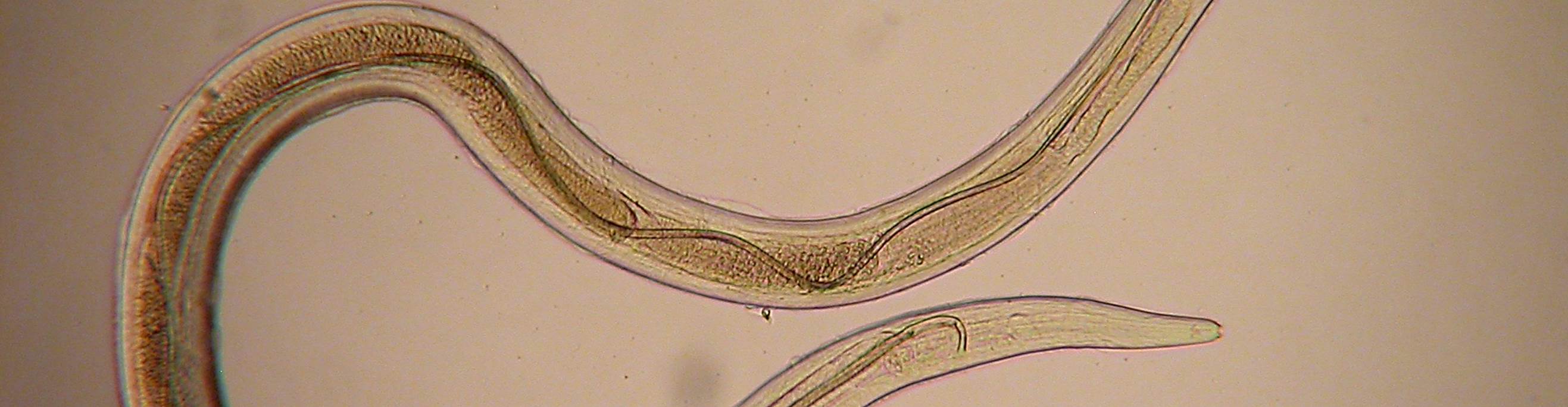
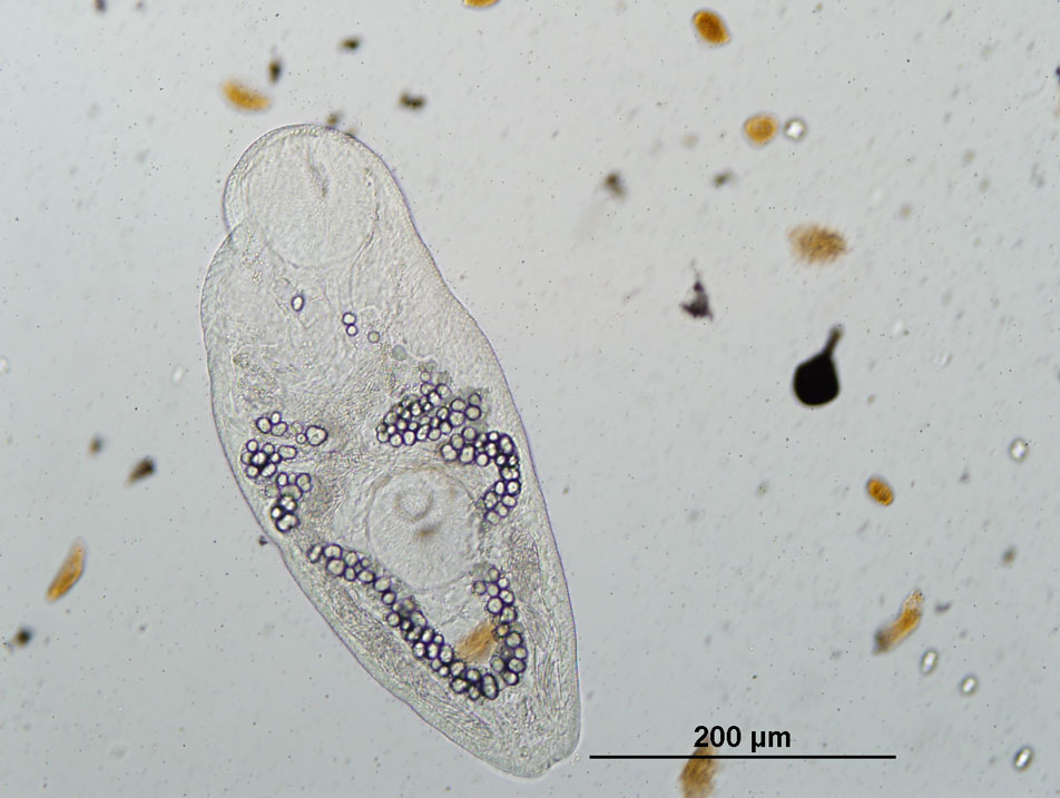
| Host Species | Host Common Name | Site(s) of Infection |
|---|
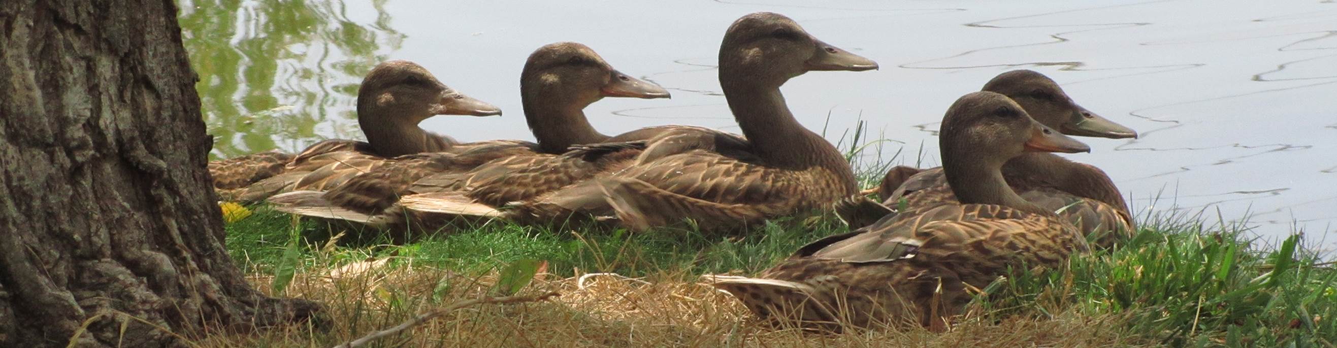
| Acris crepitans | Northern cricket frog | Skin, connective tissue, muscle |
| Ambystoma gracile | Northwestern salamander | Skin, connective tissue, muscle |
| Ambystoma laterale | Blue-spotted salamander | Skin, connective tissue, muscle |
| Ambystoma macrodactylum | Long-toed salamander | Skin, connective tissue, muscle |
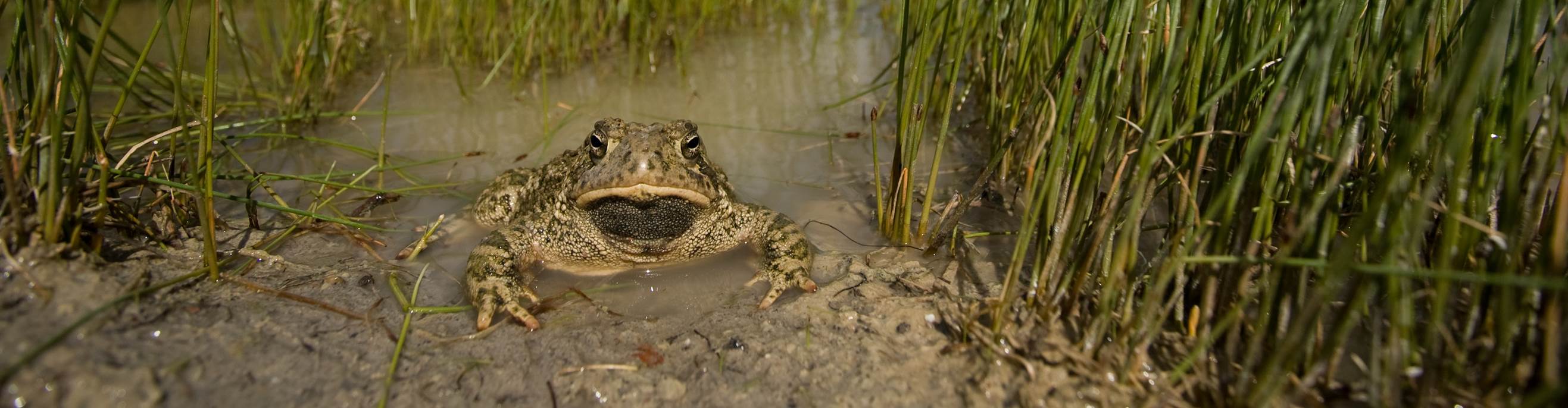
| Anaxyrus americanus | American toad | Skin, connective tissue, muscle |
| Anaxyrus boreas | Western toad | Skin, connective tissue, muscle |
| Hyla versicolor | Gray treefrog | Skin, connective tissue, muscle |
| Lepomis cyanellus | Green sunfish | Skin, connective tissue, muscle |
| Lepomis macrochirus | Bluegill | Skin, connective tissue, muscle |
| Micropterus salmoides | Largemouth bass | Skin, connective tissue, muscle |
| Pseudacris crucifer | Spring peeper | Skin, connective tissue, muscle |
| Pseudacris maculata | Boreal chorus frog | Skin, connective tissue, muscle |
| Pseudacris regilla | Pacific chorus frog | Skin, connective tissue, muscle |
| Pseudacris triseriata | Western chorus frog | Skin, connective tissue, muscle |
| Rana aurora | Northern red-legged frog | Skin, connective tissue, muscle |
| Rana blairi | Plains leopard frog | Skin, connective tissue, muscle |
| Rana boylii | Foothill yellow-legged frog | Skin, connective tissue, muscle |
| Rana cascadae | Cascades frog | Skin, connective tissue, muscle |
| Rana catesbeiana | American bullfrog | Skin, connective tissue, muscle |
| Rana clamitans | Green frog | Skin, connective tissue, muscle |
| Rana luteiventris | Columbia spotted frog | Skin, connective tissue, muscle |
| Rana palustris | Pickerel frog | Skin, connective tissue, muscle |
| Rana pipiens | Northern leopard frog | Skin, connective tissue, muscle |
| Rana pretiosa | Oregon spotted frog | Skin, connective tissue, muscle |
| Rana septentrionalis | Mink frog | Skin, connective tissue, muscle |
| Rana sphenocephala | Southern leopard frog | Skin, connective tissue, muscle |
| Rana sylvatica | Wood frog | Skin, connective tissue, muscle |
| Spea intermontana | Great basin spadefoot | Skin, connective tissue, muscle |
| Taricha granulosa | Rough-skinned newt | Skin, connective tissue, muscle |
| Taricha torosa | California newt | Skin, connective tissue, muscle |