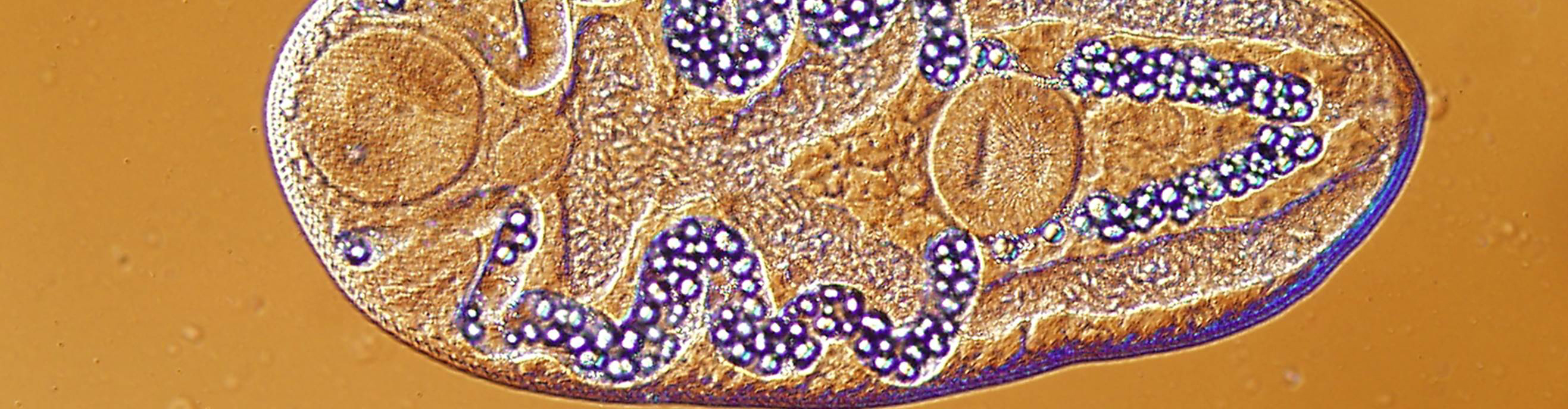
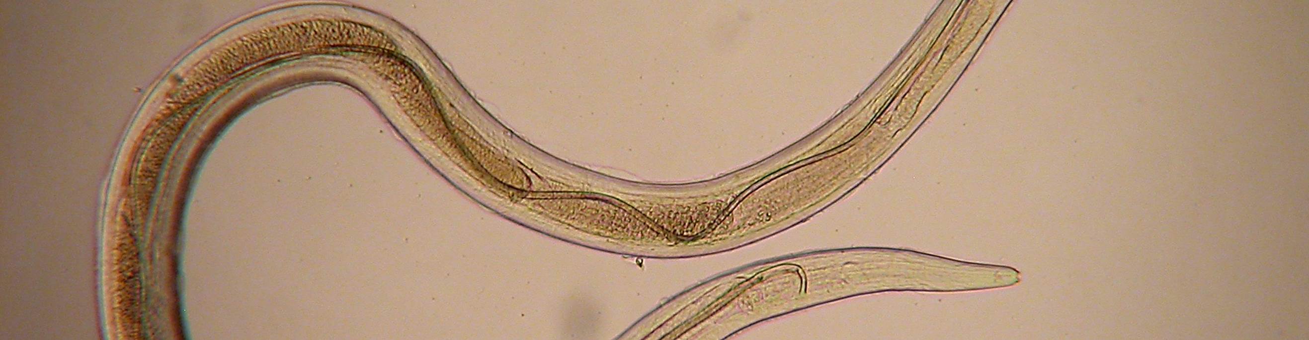
Male genitalia: Testes large and numerous and scattered through the segment both median and lateral of the longitudunal excretory canals. Vas deferens describes numerous loops near the mid-dorsal line to the anterior extremity of the segment, where it turns abruptly and enters the cirrus pouch. The cirrus pouch is in the anterior portion of the segment, is well developed and prominent, piriform, and with its posterior aperture opening alternately, usually regularly but at times irregularly, a little to the right and to the left of the median line. The cirrus from 50 microns to 1mm long, swollen at its proximal extremity and commonly found protruding in mature segments. Female genitalia: The ovaries are located in the posterior fourth of the segment, and are irregularly spherical to oval. There are two vitellaria which are partly posterior of the ovaries and partly underneath the posterior portion of the ovaries. The vagina extends anteriorly and then returns in a sinuous curve posteriorly from the genital pore and on the side of the median line opposite to the cirrus pouch; it is without a receptaculum seminis. The uterus forms as an elongate sac in the median line, and presents anteriorly a curve to one side, the cirrus pouch always lying in the concavity of this curve, and the curve being alternately, regularly or irregularly, to the right or to the left. The posterior dilation of the uterus persisting as attached cord-like structures, a short on posteriorly and a longer, sinuous one anteriorly. The eggs are ovoid, 40 to 60 microns long by 35 to 43 microns wide, and have two very thin shells."
| Host Species | Host Common Name | Site(s) of Infection |
|---|---|---|
| Acris crepitans | Northern cricket frog | Connective tissue, muscle |
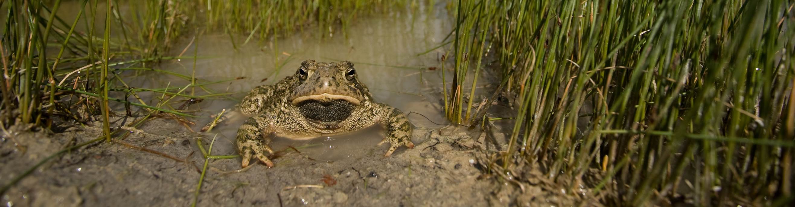
| Anaxyrus boreas | Western toad | Connective tissue, muscle |
| Anaxyrus cognatus | Great plains toad | Connective tissue, muscle |
| Hyla versicolor | Gray treefrog | Connective tissue, muscle |
| Pseudacris crucifer | Spring peeper | Connective tissue, muscle |
| Pseudacris regilla | Pacific chorus frog | Connective tissue, muscle |
| Rana catesbeiana | American bullfrog | Connective tissue, muscle |
| Rana clamitans | Green frog | Connective tissue, muscle |
| Rana palustris | Pickerel frog | Connective tissue, muscle |
| Rana pipiens | Northern leopard frog | Connective tissue, muscle |
| Rana septentrionalis | Mink frog | Connective tissue, muscle |
| Rana sphenocephala | Southern leopard frog | Connective tissue, muscle |
| Rana sylvatica | Wood frog | Connective tissue, muscle |